

Closing a surgical wound is not the end of a procedure, it is the beginning of the healing process. The choices made at the moment of closure influence tissue perfusion, scar formation, infection risk, and patient comfort for weeks afterwards. Yet suturing is often treated as the routine coda to more glamorous phases of surgery, taught briefly during training and refined haphazardly through years of clinical practice. Small mistakes accumulate, and their consequences show up in wound dehiscence, delayed healing, and the occasional call from a worried patient.
Whether you are a periodontist finishing a flap procedure or an oral surgeon closing an extraction site, mastering the fundamentals of suturing pays dividends in outcomes that patients notice. This article walks through the material selection, technique refinement, and knot-tying discipline that separate reliable closures from those that create trouble later. The goal is to help clinicians raise the standard of the final step in every procedure, without introducing complexity that slows the surgical day.
Selecting the Right Suture for the Tissue
Not every wound calls for the same thread. Absorbable materials such as polyglactin dissolve over weeks and suit intraoral sites where suture removal is impractical, while non-absorbable options such as polypropylene provide durable support in areas that need longer closure integrity. Diameter matters too, with finer threads producing less tissue reaction in delicate mucosa and heavier gauges reserved for regions under mechanical stress. Choosing poorly at this stage sets up problems that no amount of technique can compensate for.
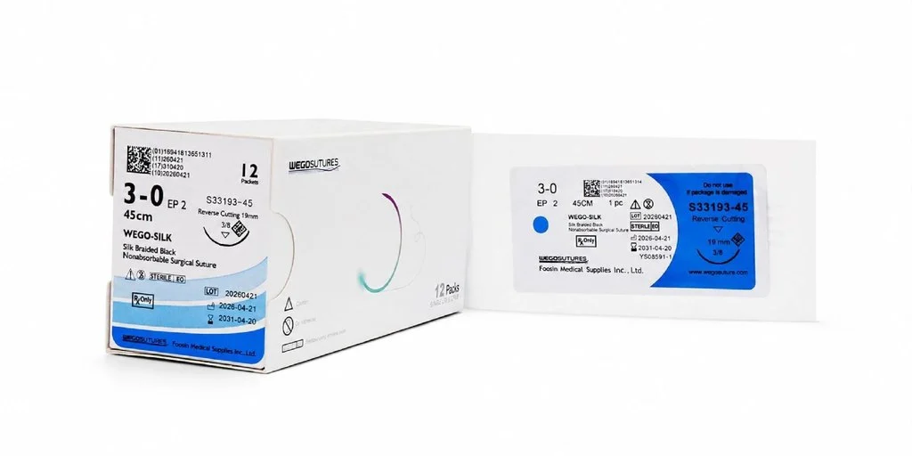
A well-stocked surgical suture inventory should include a range of gauges, needle curvatures, and absorbable profiles matched to the procedures your practice performs most often. Rather than ordering a single generic option and forcing it to fit every situation, take time to inventory the last month of procedures and identify which combinations recur most frequently. This granular approach eliminates the frustration of pulling the wrong pack from the drawer in the middle of a closure.
Technique Fundamentals That Change Outcomes
Even with ideal materials, the way the thread is placed determines how well the wound heals. Bites that are too shallow tear through mucosa within days. Bites that are too deep strangulate the tissue and impair blood supply. The classic recommendation of placing the needle roughly the same distance from the wound edge as the thickness of the tissue offers a reliable starting point that can be adjusted for site-specific anatomy.
Interrupted vs Continuous Patterns
Interrupted stitches remain the workhorse of oral surgery closure because a single failed knot does not compromise the entire line. Continuous patterns close faster and distribute tension more evenly, but they require careful anchoring at each end. New surgeons should master interrupted technique first, then add continuous patterns to their repertoire as their knot security improves. Neither approach is universally superior, and the best clinicians choose based on wound geometry rather than personal preference.
Knot Tying and Tension Management
A poorly tied knot loosens within hours regardless of how well the tissue was approximated. Square knots with three or more throws remain the standard for most intraoral applications, though monofilament materials often require additional throws because of their lower friction coefficient. Tension is the other half of the equation. Too tight, and the tissue blanches and necroses. Too loose, and the wound edges separate and permit contamination. Aim for approximation without compression, and check the colour of the tissue immediately after each knot.
Preventing Common Closure Complications
Wound dehiscence, infection, and delayed healing account for most post-surgical complaints in oral surgery practices. Each has closure-related causes that can be reduced with attention at the moment of suturing. Excessive tension is the leading cause of dehiscence, particularly in mucogingival procedures where flap mobility must be adequate before closure begins. Releasing periosteal tension before placing sutures often eliminates the problem entirely and takes only a minute of extra dissection.
Infection risk correlates with the number of suture ends left in the mouth and the reactivity of the material chosen. Trimming ends short reduces plaque accumulation, and using materials with monofilament construction rather than braided threads reduces bacterial wicking along the suture path. Suppliers such as Plovio offer suture options engineered specifically for the oral environment, where saliva exposure and mechanical challenge from the tongue create conditions that skin sutures were never designed to withstand.
Building Consistency Across the Team
In a solo practice, closure technique tends to converge on the operator’s preferences over time. In a group setting or teaching environment, however, variation between clinicians can lead to inconsistent healing outcomes and confused post-operative instructions. Establishing a short closure protocol document, with the practice’s preferred materials, patterns, and tension guidelines, gives new team members a clear reference and improves the consistency of results across chairs.
Regular case reviews focused specifically on closure quality also help. Photographing wounds immediately after suturing and again at the one-week check gives the team visual evidence of what works and what does not. Over months, this feedback loop refines technique across the group and identifies practitioners whose closures consistently produce superior healing, providing informal mentorship opportunities that formal training rarely delivers.
Closing Well, Healing Better
Suturing may be the final act of a procedure, but its influence extends far beyond the chair. Material selection, needle placement, pattern choice, and knot security combine to determine whether a wound heals uneventfully or generates the kind of complication that erodes patient trust and consumes practice hours. The good news is that each of these elements is within the clinician’s direct control, and improvement compounds quickly once the fundamentals are addressed.
Invest time in inventorying the sutures your practice actually uses, in refining technique through deliberate practice, and in building a shared standard across the team. The rewards show up in fewer follow-up complications, calmer patients, and a quiet confidence in every closure. Treat the final stitch with the same care as the incision, and the entire procedure gains a stability that makes every subsequent step easier for everyone involved.
